Referral for surgery varies slightly on patient condition but in general it is about 5 cm for women and 5.4cm for men. If the aneurysm has grown 1cm or more over the past year, this is another indication.
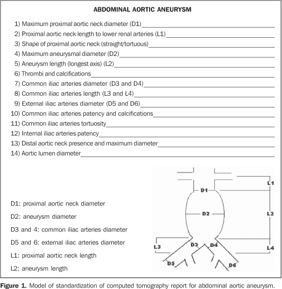
Pre-operative imaging should evaluate and document key aneurysm morphology, access vessel size, and patency including:
- Aortic tortuosity
- The proximal and distal landing zones. Ideally, these should be a minimum of 2 to 3 cm apart [6] to ensure an adequate seal and decreased rates of endoleaks, aneurysmal degeneration, and device migration.
- The proximal and distal landing dimensions as well as the aortic diameters over the graft (to determine endograft sizing).
- In the abdomen, care must be taken to assess for possible stent coverage of major branch vessels. If abdominal visceral branches are involved, and there is potential for the celiac trunk or superior mesenteric artery to be covered by the stent graft, then the presence of collateral vessels must be documented. In the absence of collaterals, an open surgical or hybrid approach may be necessary to avoid visceral ischemia [6].
- When the distal landing zone is located within one or both of the common iliac arteries, the diameter and extent must be documented.
- For conventional endovascular repair to have an adequate proximal graft seal, an aneurysm neck size of >10 to 15 mm in length and <30 mm in diameter. Over 50% of patients have aneurysm morphology unsuitable for conventional endovascular repair. Unfavorable neck anatomy, based on its diameter, length, angulation, morphology, and presence of calcification, is the most frequent cause of exclusion.
- Mural thrombus and atherosclerotic calcification covering more than 90° of the circumference of the aortic diameter in the proximal neck is associated with a higher endoleak and stent-graft migration risk.
- It is also necessary to evaluate the access path from the femoral artery through the iliofemoral vasculature [6]:
- The minimal external iliac artery intraluminal diameter should be ≥7 mm to safely accept AAA delivery sheaths.
- Since thoracic aorta endografts are larger than their abdominal counterparts, their insertion sheaths can have outer diameters up to 27 French and require a minimum vessel diameter of at least 8-9 mm.
Increased vessel depth, degree of femoral artery calcification, and iliofemoral tortuosity are negative predictors of percutaneous repair success.
Dissection:
Aortic dissection may be classified according to either the Stanford and DeBakey systems. Stanford is more widely used for TAAs; it classifies dissections into those that involve the ascending aorta as type A, and all others distal to the left subclavian artery as type B. The DeBakey system may be used for both TAAs and AAA; this system classifies dissection based on the site of origin and is divided into types I, II, IIIa, and IIIb.
The majority of dissections arise from ascending aorta – either first few cm or just distal to the origin of left subclavian. On pre-operative imaging, in addition to identifying the start and end locations, the following morphology should be described:
- Aortic dilatation
- Dissection flap (linear mobile structure within the aorta which moves more than the aortic wall)
As well as flow (Color Doppler or multi-phase imaging) characteristics:
- Flow in true and false lumens will be different and may be able to localize entry and exit points.
- There may be thrombus in false lumen (partial or no flow)
And finally, look for complications of dissection:
- Aortic rupture
EPM AAA Solution
Eon offers the most powerful solution to identify incidental AAAs, automate screening for eligible populations, and longitudinally track patients who need serial surveillance.